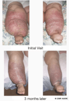
How To Assess Lymphedema
Lymphedema can be defined as swelling of one or more limbs which may also include a portion of the corresponding trunk. Lymphedema can also affect the breast, head, neck or genitalia. It occurs when fluid and other components such as protein accumulate in the tissue spaces as a result of a disparity between the creation of interstitial fluid production and its transport or movement. It may be caused by damage to the lymphatic system (i.e., as a result of cancer treatment) or as the result of congenital malformation of the lymphatic system. It is a chronic condition with no cure, but it can be managed if diagnosed early on.
General Assessment of Lymphedema
Assessment of the patient with lymphedema should be structured and ongoing. The following information should be obtained:
- extent, location and duration of the edema
- presence of lymphadenopathy (swollen lymph nodes)
- involvement/quality of skin and underlying tissue
- degree of shape distortion of the affected area
- circumference and volume of the affected limb
- presence of pitting (indicates excess interstitial fluid)- press firmly for 10 seconds with your finger or thumb for at least 10 seconds, being careful not to cause pain; if an indentation remains after you release pressure, pitting is present. The depth of any resulting indentation can be measured to assess the degree of pitting present.
Measuring Limb Volume
Limb volume measurement can be used to determine the extent and severity of lymphedema. It may also be used to assess whether treatment is effective or not. Limb volume should be assessed at the first visit, after two weeks of multilayer lymphedema bandaging and at follow-up.
When one limb is affected, both the affected and unaffected limbs are measured- the difference between the two is recorded as a percentage or in milliliters (ml). A diagnosis of lymphedema can be made if the volume of the swollen limb is 10% or more than that of the unaffected limb. When both limbs are affected, both are measured in order to track the progress of treatment. [Note: there is no effective way to measure edema affecting the head, genitalia, breast, neck or trunk. Obtaining digital photographs is recommended to record and monitor edema in these areas].
Water Plethysmography (Water Displacement Method)
The water displacement method is the gold standard for measuring limb volume and is the only reliable method available to assess edema of the hands and feet. This method follows the scientific principle that an object will displace its own volume of water. However, its use is limited by hygiene issues and ability to access this modality.
Circumferential Measurement
This is the most widely used method of calculating volume. It is reliable when a standard protocol is adhered to. Circumferential measurements of limbs can be entered into a computer program (such as a spreadsheet) designed to determine both individual and excess limb volume.
Perometry
This method uses ultraviolet light beams to measure the limb outline. Limb volume can be calculated from these measurements. The cost of the equipment limits its use, as does its inability to be used for the measurement of hand and foot volume measurements.
Bioimpedence
This method measures tissue resistance to an electrical current to measure fluid volume. A drawback to this method is that it is not very useful when bilateral swelling is present. Again, this technique is not widely used as the cost of the equipment can be prohibitive.
Limitations of Limb Volume Measurement
Measurement of limb volume is not particularly useful when both limbs are affected; however, measurements can be used to assess the effectiveness of treatment rendered. Also, limb volume measurements are less accurate in patients who have severe thickening of the outer skin layer (hyperkeratosis) or hardening of the skin with deep skin folds (elephantiasis).
Source:
Best practice for the management of lympoedema: International consensus (2006). http://ewma.org/fileadmin/user_upload/EWMA/Wound_Guidelines/Lymphoedema_Framework_Best_Practice_for_the_Management_of_Lymphoedema.pdf
Lymphedema (2013). National Cancer Institute. http://www.cancer.gov/cancertopics/pdq/supportivecare/lymphedema/healthprofessional/page1

