Vascular Evaluation – Venous Insufficiency
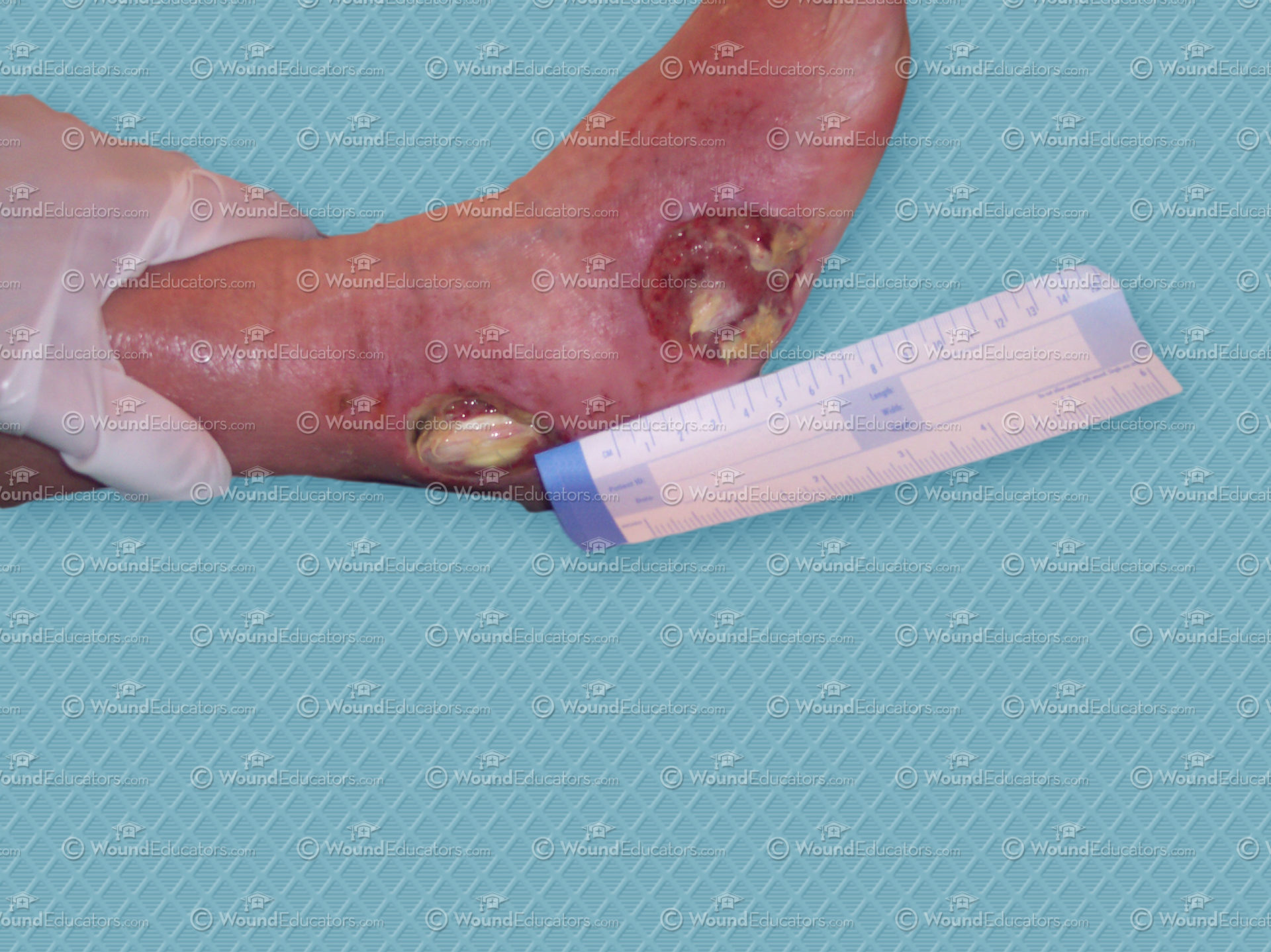
In our last article, we briefly looked at some of the techniques most commonly used in clinical practice to assess arterial insufficiency. This week, we turn our attention to venous insufficiency. Many of the techniques used are the same or similar but offer a different diagnosis and, importantly, mandate a different wound management approach.
Ankle-Brachial Index (ABI)
As with arterial insufficiency, ankle-brachial index (ABI) is commonly used in patients with suspected venous insufficiency. If the ABI is found to be below 0.8, a routine referral to a vascular surgeon should be initiated. However, if the ABI is below 0.5, then the patient should be referred as a matter of urgency.
Ultrasound & Vein Refilling
Doppler ultrasound is the gold standard for evaluation of the venous system and is performed as a three-part test consisting of resting, augmentation, and reflux. Another common and simple test of venous insufficiency is venous filling time in which the time taken for the veins to refill following elevation of the limb for one minute (or until to veins have drained) is recorded. Normal venous filling time is 5–15 seconds.
Trendelenburg Test
The Trendelenburg test may also be used to identify vein incompetence. In this procedure, the patient should be placed in the supine position with the leg elevated at 45° for one minute. A tourniquet should be secured around the distal thigh before the patient is asked to stand upright for one minute. The time required for superficial vein distention is then recorded. Distention occurring within less than 20 seconds indicates deep or perforating vein incompetence. The procedure should then be repeated without use of the tourniquet.
As in arterial insufficiency, plethysmography is used in the diagnosis of venous insufficency and is used to record changes in the volumes and sizes of extremities by measuring changes in blood volume.
Of all the methods used for assessing venous blood flow, the most accurate is actually a technique known as venography. However, as this involves injecting radiopaque dye into veins, it does not form part of routine practice.
Learn More With Our Wound Care Education Options
Interested in learning more about wound care and certification? Browse through our wound care certification courses for information on our comprehensive range of education options to suit healthcare professionals across the full spectrum of qualifications and experience.
References
- Sussman C and Bates-Jensen B. Wound Care. A collaborative practice manual for health professionals. 3rd Ed. Wolters Kluwer/Lippincott Williams & Wilkins, Philidelphia, US. 2007.
- Myers BA. The Wound Care Practitioner. Taken from Wound Management: Principles and Practice 3rd ed. Upper Saddle River, NJ: Pearson; 2012.